Friday, November 26, 2010

Radial Forearm Free Flap for Total Lower Lip Defects


Exposure to carcinogens can cause cancers of the lower lip. If these cancers are not addressed in a timely fashion, the cancer can replace the substance of the lower lip almost entirely. Surgical removal of these tumors requires total lower lip excision and bilateral neck dissection.


Large defects such as this one can not be closed primarily, by bringing the two open edges of the lower lip together. In addition, it is important to restore oral lining to the inside of the mouth. A large amount of tissue that is brought into this area requires a blood supply sustain the viability of the tissue.


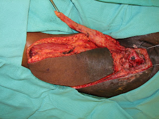
The skin, fat, and fascia on the medial aspect of the forearm is thin and pliable to conform to defects of the lower lip and oral lining. The arterial supply of the forearm is derived from the radial artery and the venous drainage is served by the cephalic vein. When incorporated properly, the palmaris longus can be used as a sling to provide oral competence.


The photograph below demonstrates from top to bottom, the cephalic vein, the radial artery, the palmaris longus tendon, and the medial antebrachial cutaneous nerve.


Appropriate inset of the flap requires microsurgical anastamosis of the flap vessels to the vesesls of the neck. The palmaris longus tendon is sutured to the modiolus and the malar eminence.  The radial artery is anastamosed to the facial artery and the cephalic vein is anastamosed to the external jugular vein.


Post-operatively, nutrition is provided by tube feeding to prevent trauma to the suture lines.


http://www.drbriandickinson.com/

Thursday, November 25, 2010

Gracilis Muscle Flap & V-Y Hamstring Flap for Closure of Ischial Defects


Defects of the trunk and lower extremity can be challenging to close. Proper closure of these wounds can allow patients to return to their activities of daily living as well as to not have to take care of an open wound. Having an open wound on the body can be difficult for patients to take care of or their family members. Patients who are in wheelchairs are prone to pressure sores. In the wheelchair patient, these sores occur on the ischial tuberosity.


Appropriate closure of these defects requires that well vascularized tissue be brought into the defect for appropriate closure. In order for the tissue to heal, it is important that all non-viable tissue be removed and for a clean wound bed to be present.  The patient must also have adequate nutritional stores. The first step in healing these wounds is proper debridement.


Once the wound is properly debrided, the next step is to cover the wound. In covering the wound, it is also important to make sure that all of the "dead space" that was created from the wound debridement is filled. The gracilis muscle is commonly used as a pedicled flap or as a free flap in microsurgery. According to Mathes and Nahai's classification, it presents a type II blood supply, based on the medial circumflex femoral artery. This artery enters the muscle about 10 cm from the pubic symphysis.
Below the gracillis muscle can be seen elevated which will be rotated and transferred into the debrided defect. Not only does the gracilis fill the "dead space" but it also provides a well vascularized tissue to deliver systemic antibiotics to any underlying osteomyelits or bone infection.


The V-Y Hanstring flap can then be elevated and advanced in the defect to close any remaining deadspace as well as to create a final wound closure.


Proper post-operative care of these wounds requires temporary immobilization to prevent shear trauma to the wound edges. Proper nutrition is also of paramount importance.


http://www.drbriandickinson.com/

Wednesday, November 17, 2010

Latisimus Dorsi Myocutaneous Flap with Extended Skin Paddle for Cross Midline Chest Wall Defects

The latissimus dorsi myocutaneous flap continues to be one of the workhorse flaps used in Plastic & Reconstructive Surgery. The words latissimus dorsi means "broadest of the back" and it is this characteristic that allows it to cover large defects locally, regionally, and for distant transfer. The latissumus dorsi muscle is a Nahai & Mathes type V muscle with its blood supply originating from the thoracodorsal vessels as well as paraspinal lumbar perforators.
I have found that the extent of the skin paddle used for the latissumus dorsi can be extended beyond those stated in standard textbooks. The skin paddle can be as wide as the breadth of the muscle and allow primary closure in the back. The length of the skin paddle can be upwards of 22 inches or greater depending upon how many perforators are incorporated into the skin paddle.
In the photo below, radiation has caused significant burn to the anterior chest leaving an open wound with exposed bone. Adequate treatment of this requires wide surgical debridement of devitalized tissue and closure with vascularized tissue. The latissimus dorsi myocutaneous flap was chosen.


The flap was marked on the posterior chest wall with demensions of 10 x 22 inches. The left of the photo marks the scapula with the right marking the iliac crest.


Dissection of the latissumus dorsi is best commenced anteriorly at the free border. Perforators are carefully coagulated as one dissects posteriorly. The muscle is then disinserted from its inferior origin along the iliac crest. The dissection around the scapula can be difficult. Thus, I find it easier to proceed from a superior to inferior direction by elevating the superior border of the muscle with double hooks and then proceeding inferiorly towards the scapula tip.


The serratus branch is then suture ligated and the pedicle is then followed proximally toward the axillae until the thoracodorsal artery and vein are identified.

The vascular leash on the latissimus is then checked to see if the muscle can be rotated freely anteriorly towards the chest wall.


A z-plasty can be completed across the axillae to allow free rotation anteriorly towards the anterior chest.


The latissimus dorsi muscle and skin paddle can extend across the midline in anterior chest wall defects.


The flap is then inset and closed over closed suction drains.

Thursday, November 4, 2010

Perforator Flap Dissection


The DIEP flap or deep inferior epigastric artery perforator flap allows the blood vessels that perfuse the skin and fat on the lower abdomen to be transferred to another part of the body. When the breast has been removed for cancer as in a mastectomy, the skin on the lower abdomen is an optimal choice to replace this missing tissue.

In athletic patients, it is helpful to preserve as much muscle as possible on the abdomen. However, it is important that the operation be done safely and to replace the missing tissue on the breast.

http://www.drbriandickinson.com/

Monday, November 1, 2010

Soleus Flap for Middle Third Lower Extremity Wounds

The Soleus Flap is used for coverage of defects of the middle third of the lower extremity. High energy wounds to the lower extremity that cause exposure of the underlying bone and fracture are best treated with coverage with this muscle.


The soleus is easily harvested via a longitudinal incision along the medial aspect of the leg. The soleus can be disinserted from the achilles tendon leaving the function of the gastrocnemius intact.


To increase the surface area of the soleus, the muscle can be scored to expand across a greater surface area of the fracture or fill in a dead space.


The photograph below shows the more superfical gastrocnemius muscle which has been left intact to preserve ankle flexion.


The soleus muscle can then be skin grafted to provide coverage of the muscle.


http://www.drbriandickinson.com/