The Flap Surgery blog is an online educational tool for patients, medical students, nurses, physician assistants, general surgery residents, plastic & reconstructive surgery residents and physicians from other medical specialties who wish to become familiar with some of the more common anatomical flaps used in plastic & reconstructive surgery.
Thursday, February 24, 2011
Zone II Flexor Tendon Injury. Repairing "No Man's Land" to Optimize Results.
Proper tendon repair to achieve maximal results with zone II flexor tendon injuries requires strict compliance of the patient with hand therapy. It is of paramount importance to adhere to post-operative care to ensure that tendons glide appropriately through the pulley system so that after the tendon heals, proper excursion can be restored to the hand to maximize outcome.
http://www.drbriandickinson.com/
Tuesday, February 8, 2011
The Sartorius Muscle Flap for Coverage of Aorto-Bifemoral Grafts
As the number of patients who are older than 65 years of age continues to gow in the United States, we are beging to see fatigue of implantable devices that may need to be replaced, exchanged, or upgraded. As a person ages, so do the vessels in the body that supply blood and oxygen to the organs. Diseases of the blood vessels that causes narrowing or weakening of the vessel wall include is called perihperal vascular disease.
The prevalence of peripheral vascular disease in the general population is 12–14%, affecting up to 20% of those over 70. Peripheral vascular disease affects 1 in 3 diabetics over the age of 50.
In the USA peripheral arterial disease affects 12–20 percent of Americans age 65 and older. Approximately 10 million Americans have PVD. Surgical treatment of peripheral vascular disease includes stenting open arteries that are narrowed or often replacing segements of arteries with grafts. Ocassionally, these grafts can become infected or exposed and necessate the coordinated efforts of Plastic & Reonstructive Surgeons with Vascular Surgeons to maximize outcomes.
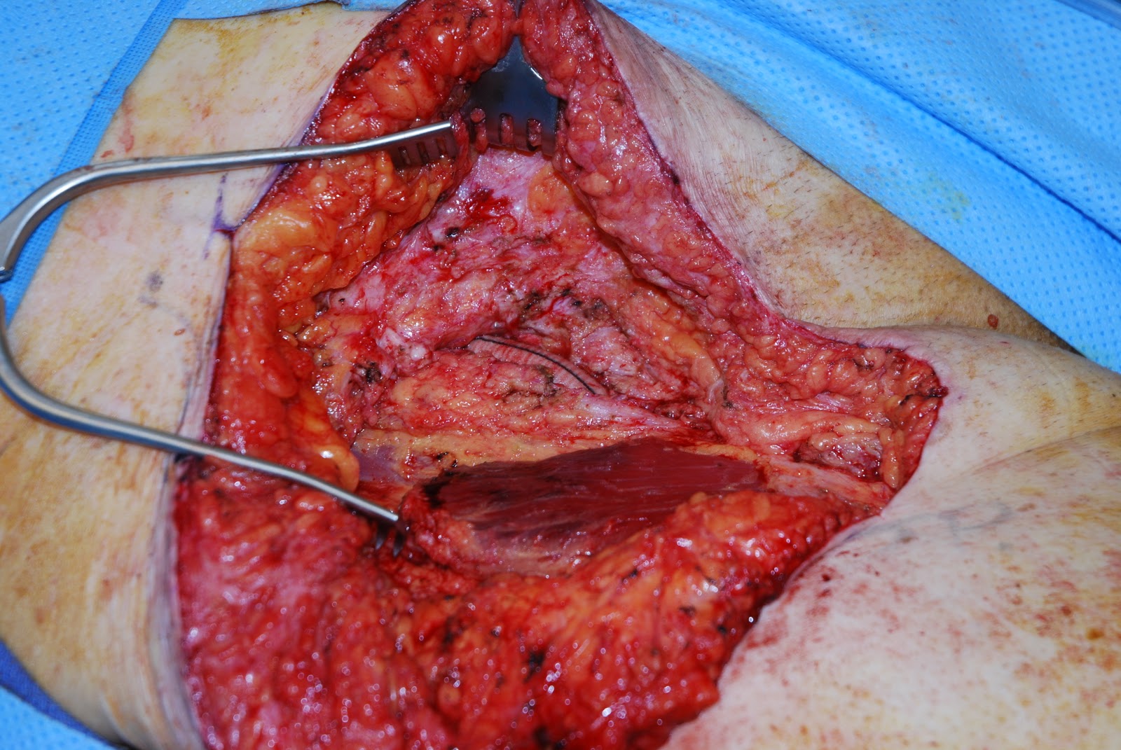
The sartorius muscle flap is a type IV muscle with a segmental blood supply. The muscle is often used in the setting of exposed, infected, or replaced bypass grafts to provided soft tissue coverage to close an anatomic "dead space' or help deliver antibiotics to the area.
The photo series below depicts a femoral limb of an aorto-bifemoral graft which has been covered with a sartorius flap to help deliver antibiotics to the region so that the graft deliver blood to perfused the leg.
http://www.drbriandickinson.com/
Thursday, January 6, 2011
Limb Salvage with Free Rectus Muscle Flap
Occlusion of vessels of the leg can significantly impair wound healing when the integrity of the skin is violated. In the distal third of the leg where there is limited leg laxity, it is necessary to bring well vascularized tissue from one region of the body to another.
When one of the three blood vessels to the leg is occluded, it is important to maintain the remaining blood supply of the lower extremity. In that case, the rectus muscle is removed from the abdomen and transferred to the leg using and end-to-side anastamosis off of the posterior tibial vessel. Using an end-to-side anastamosis can preserve the blood supply to the foot.
Drains are placed beneath the muscle and the muscle is then covered with a split thickness skin graft. Elevation of the lower extremity is important post-operatively.
http://www.drbriandickinson.com/
When one of the three blood vessels to the leg is occluded, it is important to maintain the remaining blood supply of the lower extremity. In that case, the rectus muscle is removed from the abdomen and transferred to the leg using and end-to-side anastamosis off of the posterior tibial vessel. Using an end-to-side anastamosis can preserve the blood supply to the foot.
Drains are placed beneath the muscle and the muscle is then covered with a split thickness skin graft. Elevation of the lower extremity is important post-operatively.
http://www.drbriandickinson.com/
Saturday, December 25, 2010
Axillary Flap for Closure of Chest Wall Defects
Long standing burn scars can have tumors arise in them called Marjolin's Ulcers. It is important to excise these ulcers with wide surgical margins.
Often the resulting defect that occurs can not be closed primarily and tissue needs to be borrowed from one region of the body and transferred into the defect. The axillary flap can be easily rotated into close defects on the anterior chest.
This flap allows for a better contour of the chest wall skin withou a visible or palpable depression that may occur with a skin graft.
Once the anterior chest wall wound is closed, drains are placed to evacuate any fluid from beneath the wound to prevent infection.
Post-operatively, the flap allows for adequate contour of the chest and for a closed wound.
It is not uncommon for many patients to have a wound or lesion present on their body for quite sometime. Often as these lesions are wounds grow larger, it becomes more difficult to excise, clean, or close these wounds.
What it is important for patients to know, is that there is always a solution or help that they can receive from Plastic & Reconstructive surgeons to make the wound cleaner, more manageable, or possibly close with a flap.
While the reconstructive process can often take time, patients are always happier with a closed wound that they no longer need to take care of.
http://www.drbriandickinson.com/
Thursday, December 9, 2010
Chest Wall Flap for Coverage of Exposed-Infected Elbow Hardware
Occasionally, in areas of the body where there is minimal skin laxity, orthopedic hardware can become exposed and infected. These areas where little skin laxity exists can often not tolerate the swelling associated with infection. As a result, the hardware beneath may become exposed. The Plastic & Reconstructive surgeon then borrows tissue from either an adjacent or distant site on the body to replace this tissue.
Often a VAC dressing is placed on the wound to serve as a controled suction mechanism to remove any bacteria. Once the wound is clean, a flap is performed to cover the exposed hardware and prevent infection. In this case the chest wall skin is raised and the proximal forearm wound is brought to the chest wall.
The chest wall skin is elevated based on the intercostal vessels and perforators.
The chest wall skin is then sutured to the lateral and medial aspects of the proximal forearm defect to cover the exposed hardware.
The patient remains with his arm by his side for a range of 5 to 10 days while the flap now receives its blood supply from the forearm skin. Viability of the skin paddle can be checked by placing a clamp across the chest wall flap and occluding the blood flow. If the flap remains pink, it is viable and the chest wall flap can be transferred to the forearm.
Once the chest wall flap is divided, there is brisk bleeding from the forearm skin paddle. The open medial aspect of the wound is now closed.
Finally, the expanded skin from the chest wall can be returned to successfully close the chest wall wound.
Friday, November 26, 2010
Radial Forearm Free Flap for Total Lower Lip Defects
Exposure to carcinogens can cause cancers of the lower lip. If these cancers are not addressed in a timely fashion, the cancer can replace the substance of the lower lip almost entirely. Surgical removal of these tumors requires total lower lip excision and bilateral neck dissection.
Large defects such as this one can not be closed primarily, by bringing the two open edges of the lower lip together. In addition, it is important to restore oral lining to the inside of the mouth. A large amount of tissue that is brought into this area requires a blood supply sustain the viability of the tissue.
The skin, fat, and fascia on the medial aspect of the forearm is thin and pliable to conform to defects of the lower lip and oral lining. The arterial supply of the forearm is derived from the radial artery and the venous drainage is served by the cephalic vein. When incorporated properly, the palmaris longus can be used as a sling to provide oral competence.
The photograph below demonstrates from top to bottom, the cephalic vein, the radial artery, the palmaris longus tendon, and the medial antebrachial cutaneous nerve.
Appropriate inset of the flap requires microsurgical anastamosis of the flap vessels to the vesesls of the neck. The palmaris longus tendon is sutured to the modiolus and the malar eminence. The radial artery is anastamosed to the facial artery and the cephalic vein is anastamosed to the external jugular vein.
Post-operatively, nutrition is provided by tube feeding to prevent trauma to the suture lines.
http://www.drbriandickinson.com/
Thursday, November 25, 2010
Gracilis Muscle Flap & V-Y Hamstring Flap for Closure of Ischial Defects
Defects of the trunk and lower extremity can be challenging to close. Proper closure of these wounds can allow patients to return to their activities of daily living as well as to not have to take care of an open wound. Having an open wound on the body can be difficult for patients to take care of or their family members. Patients who are in wheelchairs are prone to pressure sores. In the wheelchair patient, these sores occur on the ischial tuberosity.
Appropriate closure of these defects requires that well vascularized tissue be brought into the defect for appropriate closure. In order for the tissue to heal, it is important that all non-viable tissue be removed and for a clean wound bed to be present. The patient must also have adequate nutritional stores. The first step in healing these wounds is proper debridement.
Once the wound is properly debrided, the next step is to cover the wound. In covering the wound, it is also important to make sure that all of the "dead space" that was created from the wound debridement is filled. The gracilis muscle is commonly used as a pedicled flap or as a free flap in microsurgery. According to Mathes and Nahai's classification, it presents a type II blood supply, based on the medial circumflex femoral artery. This artery enters the muscle about 10 cm from the pubic symphysis.
Below the gracillis muscle can be seen elevated which will be rotated and transferred into the debrided defect. Not only does the gracilis fill the "dead space" but it also provides a well vascularized tissue to deliver systemic antibiotics to any underlying osteomyelits or bone infection.
The V-Y Hanstring flap can then be elevated and advanced in the defect to close any remaining deadspace as well as to create a final wound closure.
Proper post-operative care of these wounds requires temporary immobilization to prevent shear trauma to the wound edges. Proper nutrition is also of paramount importance.
http://www.drbriandickinson.com/
Subscribe to:
Posts (Atom)