Sunday, December 11, 2011

Rectus Muscle Free Flaps for Coverage of Radiated Skull Bone and Cranioplasty Plates

Superficial Temporal Artery and Vein for Recipient Vessels.

The superficial temporal vessels are excellent recipient vessels when planning for free microsurgical tissue transfer of muscle flaps to the skull. The superficial temporal vessels can be easily palpated crossing the zygomatic arch just superior to the root of the ear. The superficial temporal vessels are frequently quite toruous as one dissects proximally. It is perfectly fine to leave the vessel in it's native configuration and anastamose distal to the corkscrew of the artery.


Typically the rectus abdominis muscle provides adequate bulk and surface area to cover plates that are placed on the skull in cranioplasties. The bulk of the muslce helps to obiterate any dead space that may be present after bony debridement.


Once adequte flow is confirmed in the muscle through adequate bleeding and appropriate doppler flow, a skin graft is usually harvested from the lower extremity and then placed on top of the muscle.


www.drbriandickinson.com

Journal of Craniofacial Surgery Publication


Dickinson, Brian P.; Spoon, Daniel B.; Cordray, Tracy L.; Lazareff, Jorge; Wasson, Kristy; Bradley, James P.
Journal of Craniofacial Surgery. 17(4):707-713, July 2006.

Coverage of Massive Brain and Skull Defects with Latismus Dorsi Free Flaps

Latissimus Free Flaps for Massive Brain and Craniofacial Skull Wounds

Large defects of the craniofacial skull that are created from cancers, trauma, radiation, or other causes, often require microvascular free tissue transfer to close the wounds. On the vertex of the skull, there is limited tissue to close even small wounds and the amount of tissue is often decreased even more significantly if the tissue mobility is affected by radiation. The transfer of free muscle allows the operative surgeon to 1) cover any open wounds and 2) obliterate dead space which can often cause recurrent or recalcitrant infections.


Massive wounds of the brain and skull require complete debridement of all devitalized, radiated, necrotic, and osteomyelitic bone to allow the wound to heal properly. After all of the necrotic and devitalized material has been debrided, then the microsurgeon can bring well vascularized tissue to the area to help heal the wound.


The latissimus dorsi offers the most optimal muscle to be transferred to the scalp as it has a broad surface area to cover the entire scalp. The size match of the thoracodorsal vessels also coincide well with the size match of the superfical temporal artery and vein to allow an appropriate size match. It is important for the operative surgeon to be prepared to harvest either the greater saphenous vein or the lesser saphenous vein in the lower extremity in case the superficial temporal vein is inadequate. In this case the venous anastamosis needs to be jumped to the external jugular vein in the neck. This should be marked out pre-operatively and no central venous lines placed on that side of the neck pre-operatively.

www.drbriandickinson.com

Sunday, November 20, 2011

Free Flaps to Poplitieal Artery-Anterior Tibial Artery Saphenous Vein Bypass Grafts

As the population ages, there are an increasing number of patients who have undergone vascular bypass procedures such as popliteal to anterior tibial bypasses with saphenous vein grafts. Often, in the aging population, the only patent blood vessel to the foot is the bypass graft. When these patients incur injuries or wounds to the most distal aspect of the lower extremity, planning free flap reconstruction can be quite challenging.

I have found that in those patients, magnetic resonance angiography and magnetic resonance venograms can be helpful to establishing a correct plan for the outflow of the microvascular anastomosis. In those cases when the saphenous vein has been used, the surgeon must rely on the venae comitantes of adjacent vessels to determine successful outflow.

I have encountered, that even when native arteries have been diseased, and new blood flow has been established via bypass grafts, the venae comitantes are still often of sufficient caliber. In that case it is important for the operating surgeon to determine the location of the bypass graft tract in relation to the defect, and then select an appropriate venae comitantes in the vicinity of the bypass graft.

Either a long and sufficient caliber leash on the venae comitantes should be mobilized, in addition a flap with a long venous leash that can easily be dissected free of the artery should be selected. I have found the rectus muscle to be an excellent muscle for these cases; In that, the muscle is long and wide allowing not only for adequate coverage, but also for greater margin of separation of vascular bypass and location of nearest venae comitantes.


When the rectus has been harvested, it is important for the surgical team to spend sufficient time and care with the deep inferior epigastric artery and vein to allow adequate mobilization between both vessels.

When there is a single vessel run-off to the lower extremity, I find it helpful to perform the arterial end-to-side anastomosis first so that the flap can be rotated to-and-fro. The vein can then be usually anastomosed so that it will not be kinked. If one anastomoses the vein first, the end-to-side arterial anastomosis is often more difficult as the vein anastomosis now prevents the to-and-fro motion of the flap.

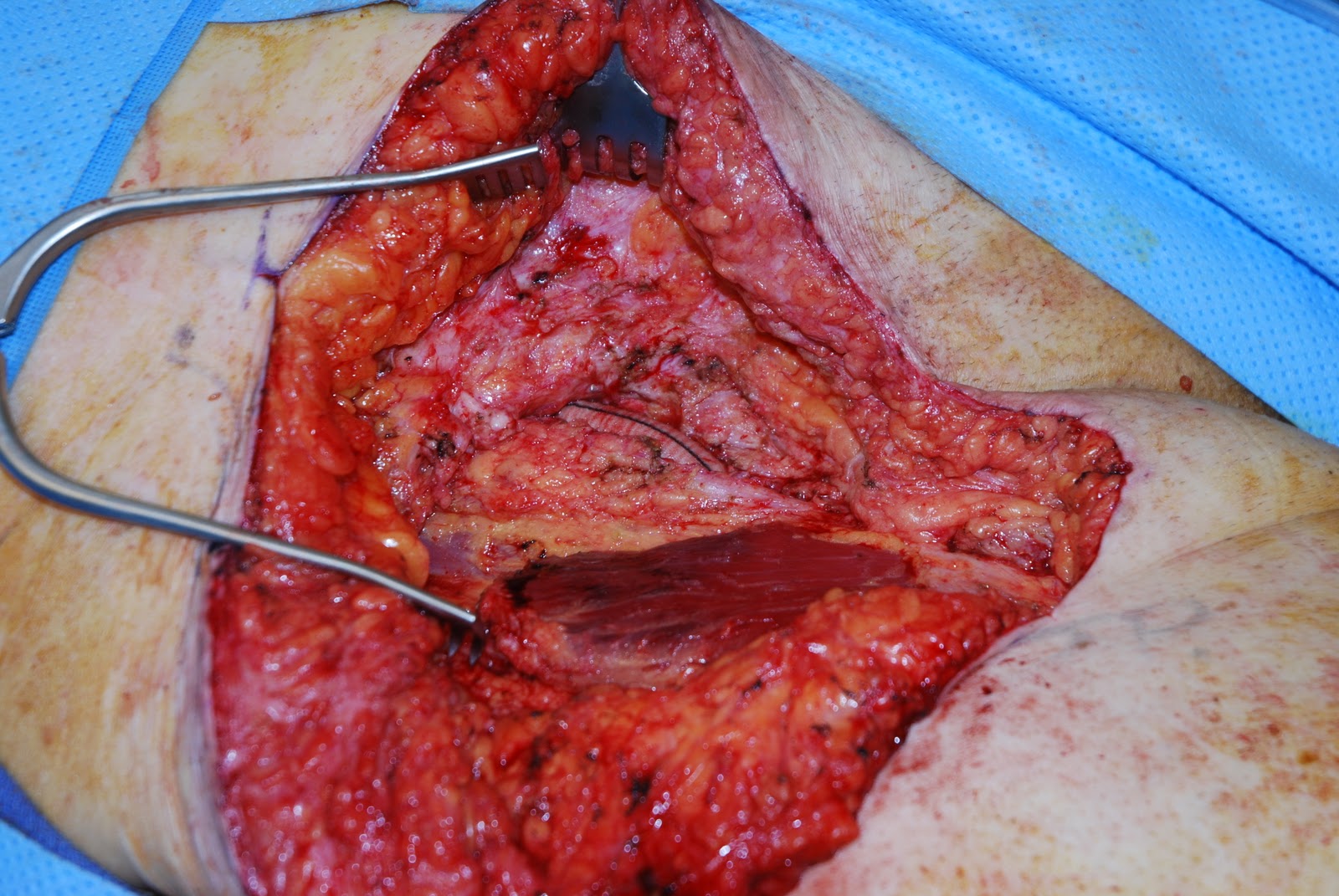
When dissecting toward arterialized vein bypass grafts, I find it helpful to gain proximal and distal control with the vascular bull-dog clamps from the vascular set. One must be aware that there is often a significant amount of scar tissue present around these grafts as they have been tunneled from their proximal to distal locations. Once adequate control has been obtained, the operative surgeons need to spend sufficient time dissecting scar off the vessel before they reach lumen.

Once the muscle has been vascularized, the split thickness skin graft can be placed.


Gracilis Free Flaps for Coverage of Achilles Tendon Wounds


It is quite common these days to see defects of the lower extremity in the region of the Achilles tendon. These wound locations are becoming increasingly more common as the population ages, is more active, and is more affected by the possibility of peripheral vascular disease.

When patients have wounds with exposed tendon it is very painful. Often wounds in the location of tendons appear to have pain out of proportion to the depth, size, and condition of surrounding skin or adjacent structures. To put it simply, wounds with exposed tendons are painful!

It is not until these wounds are covered that the patient reports relief from pain and are able to ambulate properly again.


 There are many different options for covering wounds in the distal third of the lower extremity. Depending on the location, depth, structures that are exposed, and location of quality nearby recipient vessel, I often choose among the following free flaps: 1) Rectus Abdominus, 2) Gracilis, 3) Radial Forearm, 4) Latisimus, and 5)Serratus.


The gracilis free flap offers an excellent advantage in some cases in that the surgery is confined to one surgical site and there is minimal donor site morbidity associated with the flap harvest.The gracilis muscle inserts proximally onto the pubic symphysis while the distal insertion is the medial condyle of the tibia. The blood supply of the gracilis is from the ascending branch of the medial femoral circumflex femoral artery and vein.

These blood vessels are often an appropriate size match for the posterior tibial vessels deep distal in the lower extremity.


Sunday, September 25, 2011

Free Rectus Muscle Flap to Scalp with Saphenous Vein Graft to External Jugular


Radiation is often a necessary adjuvant in the treatment of many cancers. While radiation is often necessary in cancer therapy, the radiation can also cause damage to normal healthy tissue and prevent wound healing.

When radiation is used on the head, not only can it cause injury to the skin and subcutaneous tissue, it can also cause damage to the underlying bone. When the bone develops osteoradionecrosis, it can often cause wounds to recur or simply become non-healing. When the bone is removed, often a titanium mesh or bone cement is used to cover the brain.

Usually there is not enough tissue on the scalp to close these defects, therefore free tissue transfer and microsurgery is necessary to bring in healthy tissue to the area to close the wounds.


The rectus muscle is a nice muscle to use in this scenario as there is a long pedicle leash and a muscle with a large surface area. Occasionally the superficial temporal artery is of good quality and the vein may be small. In this case we have found that the saphenous vein can be an effective vein graft from the flap to the external jugular. We find that in the past many have not advocated the use of vein grafts. However, often is more advantageous to use a vein graft to a more suitable outflow than to rely on a marginal vein in the vicinity of the wound.


www.drbriandickinson.com

Saturday, September 24, 2011

Latisimus Flap for Closure of Complex Posterior Trunk Radiation Wounds


Radiation is often a necessary adjuvant in the treatment of many cancers. While radiation is often necessary in cancer therapy, the radiation can also cause damage to normal healthy tissue and prevent wound healing.
Closure of the radiated wound is often one of the most challenging problems the Plastic & Reconstructive Surgeon can face. The key to closing radiated wounds is adequate debridement. Often adequate debridement can create a much larger wound that the original and often much larger than anticipated by both patient and surgeon.


After adequate debridement has been performed, the next important step in reconstruction is to make sure that healthy tissue is brought into the area affected to allow wound closure. In some cases, tissue from an adjacent area of the body can be used, occasionally regional tissue may be used, and in some cases distant tissue is used to be brought into the area to allow wound closure. When tissue from a remote area of the body is used, this is called free tissue transfer or microsurgical anastomosis.


In this case a large posterior trunk wound was present after successful cancer removal and successful radiation therapy. The wound was thoroughly debrided to relatively healthy tissue. A wound VAC was then placed on the posterior trunk to promote blood perfusion and as a bridge to determine the final wound size and plan reconstruction.
In this case, a latisimus myocutaneous flap was used to bring both muscle to cover exposed bone and skin to fill a defect. A small skin graft was placed on the latisimus muscle to prevent too tight of a wound closure. The skin graft however contracts over time and can often eventually be excised and the wound closed with one skin paddle.

Monday, May 2, 2011

Lower Extremity Flap Surgery. Journal of Reconstructive Microsurgery Publication

I am very fortunate to publish in the Journal of Reconstructive Microsurgery with Dr. Neil Jones on lower extremity reconstruction.

Saturday, April 9, 2011

Stacked DIEP Flap. EJ Turndown and SIEV Extension Graft


Occasionally in breast reconstruction a large portion of the abdominal flap needs to be delivered to the chest either to have significant skin surface area to replace a large defect or a large volume needs to be delivered to re-create the breast.

The stacked DIEP flap has been described to transfer a large portion of the abdominal flap to the chest wall. During my microsurgical study with Dr. Ashjian we have discovered that the venous outflow is a large determinant of flap success with stacked DIEP flaps. There is often more than sufficient arterial blood flow to sustain the volume of fat transferred when using the deep system. This is often evident when the flap is perfusing in situ on the adomen with the deep system connected and the superficial system clamped. Typically, the lateral zones develop congestion when the superficial veins are clipped. When the superficial veins are released there is often blood flow returning from the veins. If there was a lack of arterial blood flow or no connection between the deep and superficial system, then there would be no blood flow from the superficial veins.

Therefore with stacked DIEP flaps, additional venous outflow is needed to accommodate the flap volume. On the left side the venous outflow can be challenging given the traditionally smaller size of the internal mammary vein. The external jugular when mobilized and turned down into the wound can often be short depending on the body habitus of the patient. On occasion the surgeon can be presented with a patient with a short neck and long torso. This makes the reach of the EJ to the flap outflow vein challenging if the flap is already connected to the internal mammary artery. We have used the greater saphenous vein as an extension graft from the EJ to the flap. It is easier to anastomose the greater saphenous vein to the EJ extracorporeally and then pass the graft-EJ anastomosis subcutaneously into the chest.


In some situations, if the superficial epigastric veins have been preserved during flap dissection and are of adequate length, these can be used instead of the greater saphenous vein to lengthen the external jugular vein reach so that venous outflow can be augmented in the flap. This can be very helpful if both SIEVs are preserved during dissection as one can be used to anastomose to the other, offering favorable size match.


When the superficial vein is anastamosed to the external jugular vein, or other adeuate venous drainage system, with the other superficial epigastric vein as an extension graft we have found it possible to maintain almost all of the flap volume for breast reconstrution. We therefore underscore the utility of the superficial venous system in allowing venous egress of the contralateral flap and lateral flap zones.

Thursday, February 24, 2011

Zone II Flexor Tendon Injury. Repairing "No Man's Land" to Optimize Results.


Proper tendon repair to achieve maximal results with zone II flexor tendon injuries requires strict compliance of the patient with hand therapy. It is of paramount importance to adhere to post-operative care to ensure that tendons glide appropriately through the pulley system so that after the tendon heals, proper excursion can be restored to the hand to maximize outcome.

http://www.drbriandickinson.com/

Tuesday, February 8, 2011

The Sartorius Muscle Flap for Coverage of Aorto-Bifemoral Grafts

As the number of patients who are older than 65 years of age continues to gow in the United States, we are beging to see fatigue of implantable devices that may need to be replaced, exchanged, or upgraded. As  a person ages, so do the vessels in the body that supply blood and oxygen to the organs. Diseases of the blood vessels that causes narrowing or weakening of the vessel wall include is called perihperal vascular disease.

The prevalence of peripheral vascular disease in the general population is 12–14%, affecting up to 20% of those over 70. Peripheral vascular disease affects 1 in 3 diabetics over the age of 50.
In the USA peripheral arterial disease affects 12–20 percent of Americans age 65 and older. Approximately 10 million Americans have PVD.
Surgical treatment of peripheral vascular disease includes stenting open arteries that are narrowed or often replacing segements of arteries with grafts. Ocassionally, these grafts can become infected or exposed and necessate the coordinated efforts of Plastic & Reonstructive Surgeons with Vascular Surgeons to maximize outcomes.
 
The sartorius muscle flap is a type IV muscle with a segmental blood supply. The muscle is often used in the setting of exposed, infected, or replaced bypass grafts to provided soft tissue coverage to close an anatomic "dead space' or help deliver antibiotics to the area.
 
The photo series below depicts a femoral limb of an aorto-bifemoral graft which has been covered with a sartorius flap to help deliver antibiotics to the region so that the graft deliver blood to perfused the leg.


http://www.drbriandickinson.com/

Thursday, January 6, 2011

Limb Salvage with Free Rectus Muscle Flap

Occlusion of vessels of the leg can significantly impair wound healing when the integrity of the skin is violated. In the distal third of the leg where there is limited leg laxity, it is necessary to bring well vascularized tissue from one region of the body to another.


When one of the three blood vessels to the leg is occluded, it is important to maintain the remaining blood supply of the lower extremity. In that case, the rectus muscle is removed from the abdomen and transferred to the leg using and end-to-side anastamosis off of the posterior tibial vessel. Using an end-to-side anastamosis can preserve the blood supply to the foot.


Drains are placed beneath the muscle and the muscle is then covered with a split thickness skin graft. Elevation of the lower extremity is important post-operatively.

http://www.drbriandickinson.com/