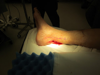
It is quite common these days to see defects of the lower extremity in the region of the Achilles tendon. These wound locations are becoming increasingly more common as the population ages, is more active, and is more affected by the possibility of peripheral vascular disease.
When patients have wounds with exposed tendon it is very painful. Often wounds in the location of tendons appear to have pain out of proportion to the depth, size, and condition of surrounding skin or adjacent structures. To put it simply, wounds with exposed tendons are painful!
It is not until these wounds are covered that the patient reports relief from pain and are able to ambulate properly again.
There are many different options for covering wounds in the distal third of the lower extremity. Depending on the location, depth, structures that are exposed, and location of quality nearby recipient vessel, I often choose among the following free flaps: 1) Rectus Abdominus, 2) Gracilis, 3) Radial Forearm, 4) Latisimus, and 5)Serratus.
The gracilis free flap offers an excellent advantage in some cases in that the surgery is confined to one surgical site and there is minimal donor site morbidity associated with the flap harvest.The gracilis muscle inserts proximally onto the pubic symphysis while the distal insertion is the medial condyle of the tibia. The blood supply of the gracilis is from the ascending branch of the medial femoral circumflex femoral artery and vein.
These blood vessels are often an appropriate size match for the posterior tibial vessels deep distal in the lower extremity.


